DermaForge® is a unique software packet for scanning and automatic analysis of patient’s moles. Using sophisticated methods of image processing and analysis developed in close cooperation with renowned dermatologists, DermaForge® offers a reliable, non-invasive and painless solution for scanning and evaluating suspicious moles on any part of the patient’s body.
The product is supplied either with a compact dermatoscope or a HEINE DELTA®20 T dermatoscope connected to a Canon digital camera.
DermaForge installation & system requirements
MS WINDOWS 10 OS
MINIMAL SCREEN RESOLUTION 1920*1080 PIXELS
MINIMUM
4GB RAM
MINIMUM 2x USB 3.0 PORTS
TOUCH SCREEN (OPTIONAL)
(DermaForge® SUPPORTS TOUCH CONTROL)
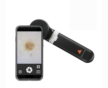
Get a fully-fledged digital dermatoscope by connecting the popular HEINE DELTA®30* dermatoscope to a Smartphone using our mobile DermaForge® VLStream application. Doctors can initiate the process of scanning simply by pressing a button on a smartphone’s screen, transferring images into the PC application DermaForge® and projecting them directly onto the PC’s screen in real-time. Pressing the button will start image analysis, images and results are stored in a database.
If a doctor already has a HEINE DELTA®30* dermatoscope, or supported smartphone they can choose to purchase the remaining parts of the set to get the functionality of a digital dermatoscope, saving the costs of a HEINE DELTA®30* dermatoscope.
Supported Smartphones and Mounting cases
| Apple iOS | Mounting case |
|---|---|
| iPhone 7/8 | Mounting case for Apple iPhone 7/8 K-000.34.261 |
| iPhone SE 2020 | Mounting case for Apple iPhone 7/8 K-000.34.261 |
| iPhone X/XS | Mounting case for Apple iPhone X/XS K-000.34.262 |
| iPhone XR | Mounting case for Apple iPhone XR K-000.34.263 |
| Google Android | Mounting case |
| Samsung S6 | Universal Smartphone Connector K-000.34.270 |
| Samsung S7 | Universal Smartphone Connector K-000.34.270 |
| Samsung S8 | Universal Smartphone Connector K-000.34.270 |
Mobile application DermaForge VLStream (DVLStream)
 |
 |
 |
 |
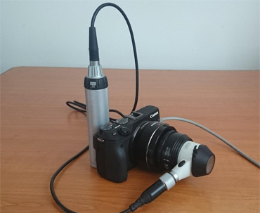
Get a fully-fledged digital dermatoscope by connecting the popular HEINE DELTA®20 T* dermatoscope to a Canon digital camera using our DermaForge® application. Doctors can initiate the process of scanning simply by pressing a camera button, transferring images into the application and projecting them directly onto the screen in real time. Pressing the camera button again will stop the scanning process automatically, whereupon the images are analysed and all images and results are stored.
If a doctor already has a dermatoscope of the type mentioned above, they can choose to purchase the remaining parts of the set to get the functionality of a digital dermatoscope, saving the costs of a HEINE DELTA®20 T* dermatoscope.
Supported Canon Camera Types
EOS-1D X Mark II
EOS 80D
EOS 1300D
EOS 5DS
EOS 5DS R
EOS 760D
EOS 750D
EOS 7D Mark II
EOS 1200D
EOS 70D
EOS 700D
EOS 100D
EOS-1D C
EOS 6D
Technical Specifications of the recommended Canon EOS 1200D/1300D
Dermatoscope: HEINE DELTA®20 T*
Adapters: HEINE Accessory Set to suit Canon Digital SLR Camera
Cables: Mini USB cable of at least 2 metres
Micro image camera: Canon EOS 1200D/1300D body
Resolution: 18 megapixels
Controlled by the shooting button located on the body of the camera
Sensor: Colour CMOS
Interface: USB 2.0
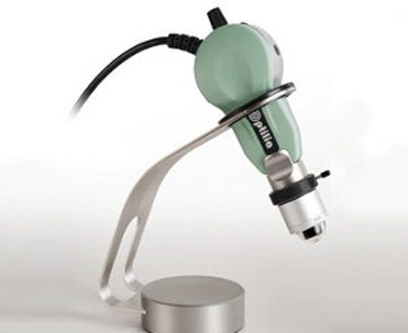
Optilia Mediscope configured for dermatoscopy offers a state-of-the-art system featuring an advanced hand-held digital camera with superior optics and light polarising illumination.
The Mediscope can be used on all areas of skin and the system of lenses provides sharp and detailed images with accurate colour representation, from 20x to 50x magnification.
Optilia Mediscope* Technical Specification
Mediscope Digital video microscope
Resolution: 3 Megapixels
20x-50x varifocal lens with LED illumination and dry polarized optics
20x dry extension contact adapter
50x dry extension contact adapter
Frame Rate: 10 fps
Illumination: 12 white ultra bright LEDs
Capture Control: Image capture foot-switch and microscope body switch
Sensor: colour CMOS
Interface: USB 2.0
Dimensions: length 14,5 cm, width (diameter) 5,5 cm
Weight: 250g (with lens)
Other components included: Aluminum transport case, Desk-top holder for Medicsope, Image capture foot-switch for Mediscope
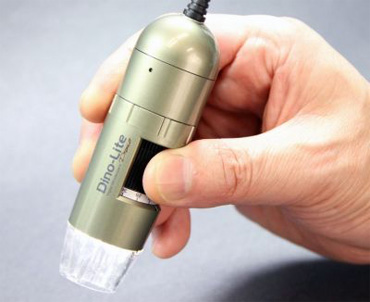
A handy and lightweight handheld digital microscope with built-in white LED lights, comfortable manual focusing and a polarizing filter for detailed skin surface scanning.
Designed for use in dermatology, the device meets all hygienic requirements and requires only basic maintenance.
DINOLITE* DERMACOPE Technical Specification
Resolution: SXGA 5 Megapixels
Zoom: 10-50x
Scanning speed: up to 30 images per second
Light: 8 built-in white LEDs (software controlled)
Polarizing filter: removable
Focusing: manual
Microtouch control button
Sensor: colour CMOS
Interface: USB 2.0
Dimensions: length 10.5cm, width (diameter) 3.2cm
Weight: 140g
Other components included: handy stand, contact cover (spare one can be ordered if damaged)